- Home
- Soo Khee Chee Surgery
Soo Khee Chee Surgery

Address
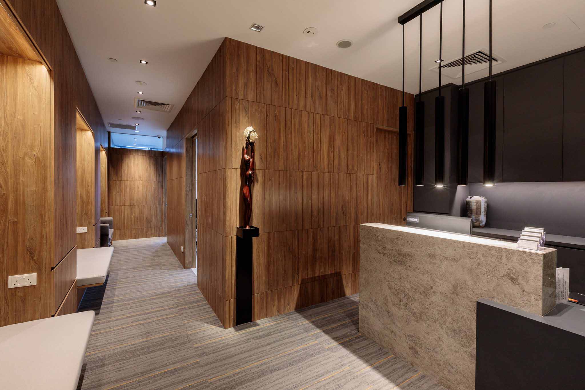
1 Farrer Park Station Road, #10-09 Connexion Farrer Park Medical Centre 217562
Phone
+65 6443 3238

Company Profile
PRACTICE AREAS
- Head and Neck Cancers
Most head and neck (H&N) of cancers are seen in the older patients, commonly those who smoke and drink. There is new trend noted of human papilloma virus related cancers seen in the younger patients with oropharyngeal (throat) cancers. The common symptoms H&N cancers are a lump in the mouth, throat, discomfort or pain, occasional bleeding with blood noticed in the saliva or enlarged neck lymph nodes. They are diagnosed after a careful H&N examination including using a flexible endoscope through the nostril to inspect the different sites in the region. Biopsies are nearly always needed. Sometimes a fine needle aspiration cytology (FNAC) can be done especially for the enlarged node(s) in the neck. Survival depends primarily on the stage of the disease and reliable treatment. The majority of cases should be discussed in the H&N tumour boards though in some the appropriate treatment is obvious based on international guideline commonly the National Comprehensive Cancer Network (NCCN) guidelines.
In general, in early stage disease (stages 1 & 2) the treatment is by surgery removing the primary tumours. Radiation therapy is however the treatment of choice for tumours of the voice box as the intent is to cure but also to preserve speech. We achieve control of the disease in a significant number (up to 90% in early laryngeal cancers). Even in some patients that have failed radiation therapy we are able to do salvage surgery conserving the larynx (Figs 1, 2, 3, 4 and 5). In the advanced stages (3 & 4) the treatment usually consists of surgery followed by postoperative radiation with or without chemotherapy. Immunotherapy or photodynamic therapy may be used for recurrent unresectable tumours.
- Peritoneal Malignancies
The abdominal and pelvic cavities together the peritoneal cavity. Intraabdominal tumours breaking through their radial margins will shed tumour cells into the peritoneal cavity. This is commonly seen in ovarian, colorectal, gastric and other abdominal cancers. There are also some rare tumours like primary peritoneal cancers, peritoneal mesothelioma and pseudomyxoma peritonei. In all of these, previously deemed incurable, we now have a surgical technique known as cytoreductive surgery and HIPEC where in correctly selected patients with no evidence of distant or visceral metastasis, we will attempt to remove all the peritoneal deposits (cytoreductive surgery) and follow up intraoperatively with high dose intraperitoneal chemotherapy delivered at 40 C (HIPEC) (Fig 1 and 2). We pioneered this operation in Singapore more than 15 years ago. Our results in a highly selected group of patients have shown good survival rates compared to historical controls (Fig 3).
- Sarcomas
Sarcomas are cancers of the musculoskeletal system and the connective tissues. They are rare tumours and the management is best discussed in a multidisciplinary team. In general there are bone, soft tissue, intra-abdominal and retroperitoneal, pelvic, chest and head and neck sarcomas. While some sarcomas may need neoadjuvant (upfront) chemotherapy, for most the treatment consists of definitive surgical resection with or without postoperative radiation (Figs 1, 2, 3 and 4). (A compartmental resection). The ability to achieve clear surgical margins is critical. Sometimes preoperative radiation therapy is used to downstage the disease to facilitate surgery and also in the postoperative setting to reduce local recurrence. Chemotherapy is used for recurrent or metastatic disease.
The prognosis is dependent on the size of the primary, the grading of the tumour, the tumour type (histological classification) and the completeness of surgical resection.
- Thyroid
Most patients with thyroid goitres do not need surgery. The indications for surgery are if the goitre is so large that it is compressing on the airway or affecting swallowing (Fig 1), or there is suspicion of cancer or proven to have cancer on a fine needle cytology (FNAC). Sometimes toxic goitres are surgically removed if the thyrotoxicosis is not well controlled medically. On rarer occasions some goitres are removed for reason of cosmetics.
All goitres will need to be assessed with thyroid function test, an ultrasound and for suspicious or obvious nodules with flab. Sometimes FNAC (Fig 2) are not definitive especially for patients with reported follicular lesions and in such situations we would recommend surgical removal.
Thyroid cancers are of 4 main types. The commonest and the one with best prognosis is papillary cancer. This occurs in the younger age group (30-40 years). The others are follicular, anaplastic and medullary cancers. Anaplastic cancer stands out for being one of the most aggressive cancers in the human boy. Fortunately they are infrequent, commonly see in elderly patients with fast growing thyroid goitres. Most thyroid cancers are treated with a total thyroidectomy (Fig 3 and 4). In this operation great effort is made not to damage the two recurrent laryngeal nerves which lie just behind the thyroid gland. Care is also taken not to remove or damage the 4 parathyroid glands. These control calcium metabolism. In the postoperative setting some patients will require radioactive iodine ablation. If so they will need to be admitted to the hospital as their body fluids will be radioactive for about 3 days.
- Salivary Glands
We have 3 paired major salivary glands – the parotid, sublingual and submandibular glands. We also have thousands of small minor salivary glands in the upper aerodigestive tract.
The large majority of tumours are in the parotid gland (70%) followed by the submandibular gland (8%) . 60% of salivary gland tumours are benign. Most salivary gland tumours will need to be confirmed by CT or MRI imaging. Fine needle aspirate cytology (FNAC) is done for the majority though a strong argument can be made for not doing it routinely as the majority of the tumours will need surgical excision in any case. In my practice, I will do FNAC in patients in whom I am wanting to avoid surgery e.g benign tumours in the elderly, or those where I am suspecting the swelling is due to a lymphoma.
Patients with tumours of the parotid gland will require a formal superficial parotidectomy. In the majority of cases, 90% of parotid tumours are found superficial to the facial nerve which divides the parotid salivary gland into a superficial and deep lobes. The operation of superficial parotidectomy preserves all the 5 branches of the facial nerve running through the gland and this is important because these branches provide the motor innervation to the side of the face (fig 1,2,3). However, in about 25% of the patients even with the careful preservation of these 5 branches, there is facial weakness most of which will go away after 2 to 3 months. It is unusual to require sacrifice of the facial nerve in parotid tumours unless if the face is already paralysed. Some of the tumours may require concurrent lymph node dissection and in others additional postoperative radiotherapy.
- Advanced Abdominal Malignancies
Patients with intraabdominal cancers are often those who have recurrent disease or those who have late presentations because these tumours are deep seated. While the prognosis worsens with recurrence or late presentation, there are situations where surgical salvage is possible. Many factors need to be considered including:
1. Whether there is distant spread outside the abdominal cavity to the lungs, brain etc
2. Invasion of adjacent critical structures e.g blood supply to the length of the small intestine
3. The biological behavior of the tumour ie rapidity of the return of disease, the number of lymph nodes involved radiologically
Careful consideration of these factors and others will help determine the possibility of surgical resection. If surgical salvage is possible with either complete removal of the disease or maximal debulking of disease, chemotherapy and radiation can be added as adjuvant treatment for additional control of disease.
Case illustration:
A 50 year old man with local recurrence after the removal of right kidney for aggressive squamous carcinoma. The tumour had extended beyond the tumour bed to the right diaphragm and right liver. The local recurrence site was removed including part of the diaphragm and the liver. Reconstruction to cover the defect was achieved with a nylon mesh and abdominal muscle flap (Fig 1 to 6). The patient remains disease free 7 years after surgical salvage.
Medical Professional(s)

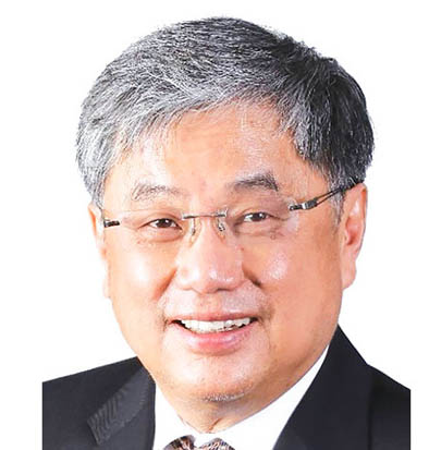
Prof. Khee Chee SOO
蘇 啟智
Surgical Oncology
Video Consult Clinic Visit
1 Farrer Park Station Road, Farrer Park Medical Centre, #09-07 Connexion, Singapore 217562
Languages: English, Chinese


