
What is magnetic resonance imaging (MRI)?
For this article, Medicus’ patient case manager had the chance to visit Quantum Medical Imaging to ask Dr Su Ann LEE on imaging technologies and how it benefits the patients and help doctors in the diagnosis of diseases. Dr. Lee is a specialist in diagnostic imaging, accredited in magnetic resonance imaging (MRI), computed tomography (CT), mammogram, bone densitometry, ultrasound and X-ray. She performs general radiology and has interests in Body as well as Neuro Imaging. Below is an excerpt from our Q&A with Dr. Lee.
What is magnetic resonance imaging (MRI)?
MRI uses a strong magnetic field to produce pictures of the body. No X-rays or ionising radiation is used, and you will not feel anything during the scan. MRI pictures are more detailed than any other medical imaging method, and can detect tumours or fractures that are too small to be seen on X-ray.
Why do doctors recommend a MRI?
If your doctor suspects an injury or disease to do with soft tissues, MR really is the imaging modality of choice to aid diagnosis. The referral letter from your doctor will help us tailor techniques and protocols for your MR scan. Just like how a Michelin starred chef would combine his best ingredients for your fine dining experience. So, it’s time to leave the fine tuning and technical bits to us and we will get you the detailed answers you want to your soft tissues.
How does a MRI help doctors in the diagnosis of diseases?
Soft tissues in our body are made up of a large proportion (about 70-90%) of water. When you sustain an injury or when an organ is diseased, the properties and amount of water in the tissue can change dramatically. This makes MRI an excellent imaging tool because it is based upon the sensitivity towards alteration in water properties in the region or organ of interest. Different tissues have different brightness on MR images. It is this image contrast that allow us to differentiate which tissue is which, including abnormal tissues. For example, in the brain, stroke is bright and normal brain tissue is gray. In the knee, a tear in the meniscus, which is the knee’s cushion, is bright and normal meniscus is black. In reality however, it is not that straightforward as to roses are red and violets are blue because there are other variables to navigate through. But you get the idea.
Is MRI safe?
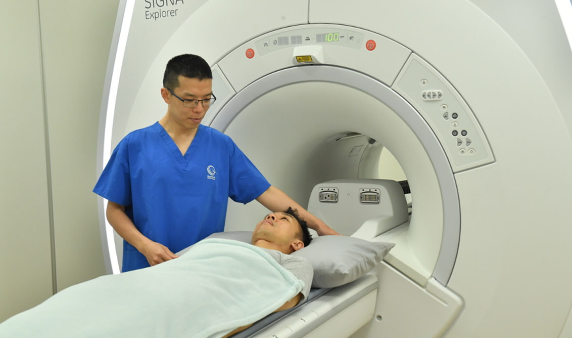
All patients will be observed during the entire exam, either through the observation window from the control room and/ or by CCTV. Our intercom will enable a 2-way audible communication between you in the scanner and the MR radiographer in the control room.
Should you be worried before a scan?
Some patients are worried before the scan. Some show signs of claustrophobia (fear of confined spaces) as the couch makes its way to the center of the scanner. These are not uncommon and to us, there is always a solution to any problem. You can be assured that our MR radiographers who take you through the scan are certified in the equipment he or she operates and have been compassionate in managing patients’ fears.
For some scans, an MR contrast agent, usually a gadolinium compound may be required so an abnormality is better defined and shows up more clearly. This involves a small injection and at Quantum, it is usually performed by the specialist on-site. In all, this group of patients comes to us nervous about the experience but leave Quantum feeling well cared for.