Vitrectomy

A vitrectomy is a microsurgical procedure done by a Vitreo-Retinal surgeon. This means that surgery is done under a microscope using miniaturized instruments by a specialist surgeon trained specifically for this delicate surgery on the back of the eye. This procedure is done for conditions like retinal detachments, advanced diabetic eye disease, epiretinal membrane, macular hole, vitreous haemorrhages (bleeding inside the eye) and complicated cataracts. It is done in an operating theatre under local or general anaesthesia.
How a vitrectomy is done
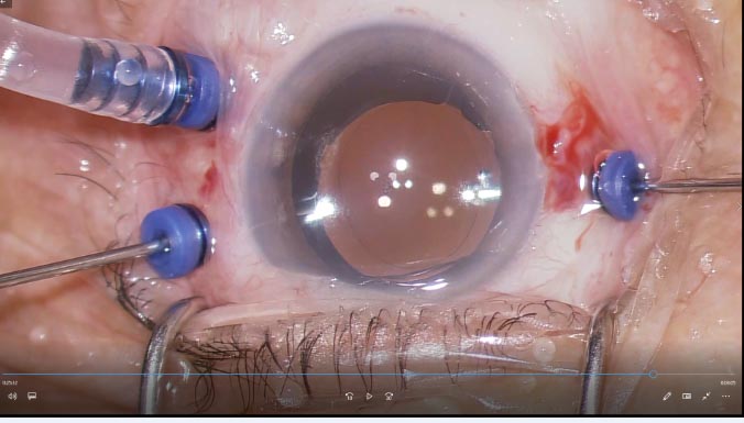
Tiny cuts are made in the white of the eye and microsurgical instruments are inserted into the back of the eye through these cuts. With the advanced sutureless technique, these cuts are very small ( 0.4 – 0.6 mm in size) and therefore require no stitching. This means that there are less pain and discomfort after surgery, and recovery is much faster than the traditional method which involves larger cuts.


The vitreous gel of the eye needs to be removed carefully before repairs can be done to the retina. Any blood and vitreous debris will also be removed.
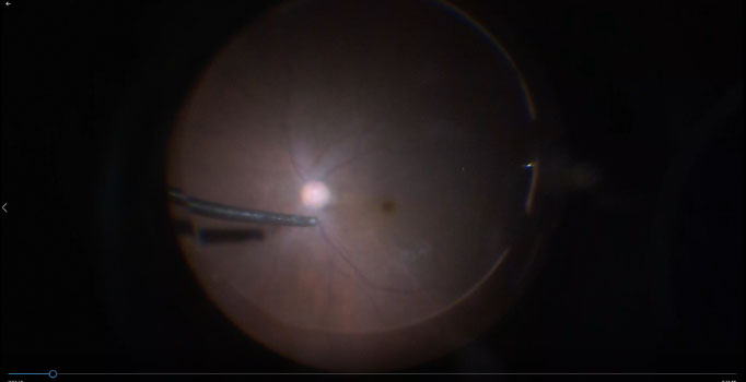
After the gel, blood and debris have been safely removed, repairs can then be done for the specific condition involved. For example, laser or cryotherapy may be needed to repair retinal detachments, or membranes may need to be peeled off using special micro-forceps.
At the end of the surgery, the eye may be filled with gas, salt solution. In some cases, it may be necessary to leave a special gas or silicone oil in the eye to help stabilize the retina. Silicone oil is used in more severe cases, or in cases where patients may need to take a flight soon after surgery. If silicone oil is used, a smaller surgery will need to be done several months later to remove this oil.

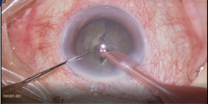
The cataract has been split into 2 halves and will be further broken up into smaller fragments to be removed.


