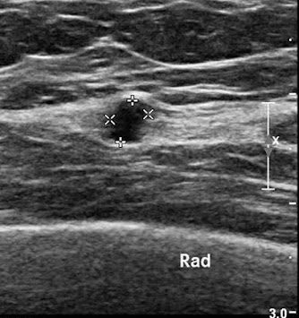
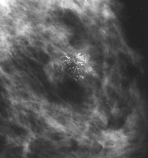
Breast screening aims to detect cancer when it is still early. Mammograms are used for this purpose. Younger women with dense breast tissue may have supplementary breast ultrasound performed as well. These 2 methods will often pick up many lumps and abnormalities. If these are biopsied and are cancerous or abnormal the woman will need to undergo surgery to have it removed.
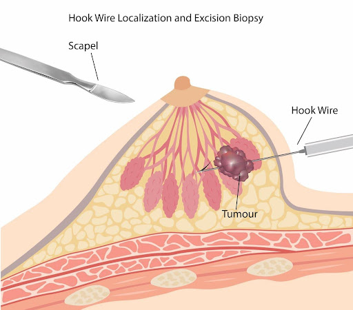
The woman arrives on the day of surgery to the hospital and under local anaesthetic a hookwire is inserted by a radiologist aiming to spear through the lump or detected abnormality. This step is sometimes performed by the surgeon in the operating room under general anaesthesia, just prior to the surgery.
Based on where the lump is in relation to the inserted hookwire, the surgeon estimates the position of the lump on the breast. She then decides where to place the incision – hiding the incision as best possible, it still needs to be close enough to remove the lump, not too long and not too short. Very often I make a periareola incision (one that is around the edge of the areola) and create a tunnel by separating the breast tissue from the skin envelope, all the way to the position of the lump. The lump is removed, together with the hookwire in the middle of it, leaving a cavity that is closed, taking great care that there are no residual pulls or dimpling on the breast skin. The incision is closed, the woman wakes up, rests for awhile and returns home the same day.
This technique is also useful when there is an abnormal larger area that needs to be removed such as a wide area of microcalcifications seen on mammogram. Instead of a single hookwire we now insert 2 to 3 hookwires that are used as borders to mark the limits of the abnormal area. The aim of the surgery is to remove the area with the hookwires in place.